EUH Morning Report: What is the reliability of the physical examination of the chest and accuracy of findings for diagnosing pleural effusion?

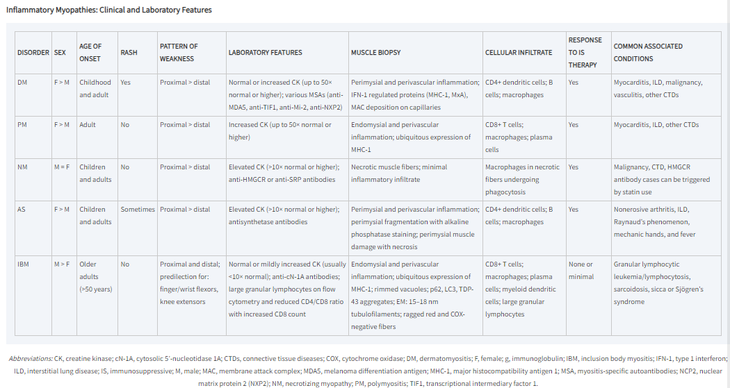
The Bottom Line: The interobserver reliability of the physical examination of the chest in patients with respiratory conditions has been found to be relatively low (Metlay). But the physical examination can be used to gain diagnostic certainty with pulmonary effusion, specifically the findings of dullness to percussion and asymmetric chest expansion, and confirmed with a chest radiograph (Wong).

“The calculated interobserver reliability among the physicians for several chest signs…  presented in the form of both mean pair observer agreement rates and κ values, which account for rates of chance agreement ranging from 0, when agreement is no better than chance, to 1, when there is perfect agreement. In fact, 2 of the most reliable findings, dullness to percussion and wheezes on auscultation, had only fair to good κ values of 0.52 and 0.51, corresponding to agreement rates of 77% and 79%, respectively. Crackles had a κ value of 0.41 (agreement rate of 72%), and several findings such as whispered pectoriloquy and increased tactile fremitus had κ values indicating poor agreement (range, 0.01-0.11), in part explained by the rarity of these findings overall.” (Matley)

(Matley)

“Of the 8 physical examination maneuvers, the presence of dullness to conventional percussion (summary positive LR, 8.7; 95% CI, 2.2-34) and asymmetric chest expansion (positive LR, 8.1; 95% CI, 5.2-13) were most accurate in diagnosing pleural effusion. The diagnostic OR of the 2 studies that compared conventional percussion (summary diagnostic OR, 34; 95% CI, 16-72) with auscultatory percussion (summary diagnostic OR, 8.1; 95% CI, 4.7-14.0) favored conventional percussion. The extremely low negative LR for auscultatory percussion popularized by Guarino (negative LR, 0.05; 95% CI, 0.02-0.11) has not been replicated in other studies (negative LR range, 0.50-1.0).” (Wong)

Continue reading

EUH Morning Report: Review of Mitochondrial Diseases

The Bottom Line: “Most diagnostic algorithms [for mitochondrial diseases] recommend evaluation of selected mitochondrial biomarkers in blood, urine, and spinal fluid. These typically include measurements of lactate and pyruvate in plasma and cerebrospinal fluid (CSF), plasma, urine, and CSF amino acids, plasma acylcarnitines, and urine organic acids.” (Parikh)

“The key to any successful diagnostic algorithm for mitochondrial diseases is astute clinical observation and awareness. The recognition of mitochondrial disease syndromes or specific clinical features can permit targeted genetic analysis, which enables the rapid diagnosis of patients and family members. For example, cardiomyopathy and cataracts are frequently associated with AGK mutations (Sengers syndrome). Equally, detailed clinical and laboratory tests are vital for the accurate interpretation of new genetic mutations that are identified through next generation sequencing technologies. The diagnostic algorithm is heavily influenced by the age of the patient, and the presence of consanguinity and common genetic founder mutations in certain parts of the world.” (Gorman)

(Gorman)
Continue reading

EUH Morning Report: What are the diagnostic accuracy of the Kernig and Brudzinski sings for diagnosing adult meningitis? Part 2

The Bottom Line: “In the broad spectrum of adults with suspected meningitis, 3 classic meningeal signs did not have diagnostic value; better bedside diagnostic signs are needed.” (Thomas)

See Part 1 on the Kernig and Brudzinski signs: https://emorymedicine.wordpress.com/2024/03/14/euh-morning-report-what-are-the-sensitivity-and-specificity-of-the-kernig-and-brudzinski-sings-for-diagnosing-adult-meningitis/

Thomas et. al’s prospective study found that “the 3 classic meningeal signs—Kernig’s sign, Brudzinski’s sign, and nuchal rigidity—were of limited clinical diagnostic value for adults with suspected meningitis. None of these meningeal signs were able to accurately discriminate patients with meningitis (⩾6 WBCs/mL of CSF) from those without it. Furthermore, no significant correlation existed between these meningeal signs and moderate meningeal inflammation (⩾100 WBCs/mL of CSF) or between these meningeal signs and microbiological evidence of CSF infection. Only for the 4 patients with severe meningeal inflammation (⩾1000 WBCs/mL of CSF) did nuchal rigidity have 100% sensitivity, 100% negative predictive value, and LR+ : LR- that approached infinity.” (Thomas)

“The sensitivity of both Kernig’s sign and Brudzinski’s sign was 5%, which suggests that these bedside diagnostic tools did not reliably identify the need for lumbar puncture among patients with meningitis. Although the specificity of both signs was 95%, the high specificity values were a result of the overall paucity of positive results of examination for Kernig’s sign and Brudzinski’s sign, rather than a reflection of the discriminating ability of these indicators. The positive and negative predictive values for Kernig’s sign (27% and 72%, respectively), Brudzinski’s sign (27% and 72%, respectively), and nuchal rigidity (26% and 73%, respectively) also indicate that none of the classic meningeal signs were clinically discriminating indicators of the presence or absence of meningitis” (Thomas)

(Thomas)
Continue reading

EUH Morning Report: What are the important clinical features for diagnosing the different types of inflammatory myopathies?

The Bottom Line: “Idiopathic inflammatory myopathy (IIM) includes dermatomyositis (DM), polymyositis (PM), overlap myositis (OM), sporadic inclusion body myositis (IBM) and necrotising autoimmune myopathy (NAM), also known as immune-mediated necrotising myopathy. DM, OM and NAM all present similarly, with proximal weakness and elevated creatine kinase (CK) level. By contrast, IBM preferentially involves the long finger flexors and quadriceps, and presents with a normal or only mildly elevated CK. Developments in serological testing and imaging are shifting the diagnostic paradigm away from a reliance on histopathology.” (Ashton)

“There is some debate as to whether PM exists as a discrete entity, or is an ill-defined condition encompassing connective tissue disease (CTD) associated myositis, or OM, and the previously poorly recognised NAM” (Ashton)

(Greenberg & Amato)

“Epidemiologic studies suggest that the incidence of IM grouped together is >4 cases per 100,000 with prevalence in the range of 14–32 per 100,000. Defining the actual incidence and prevalence of the individual myositides is limited, however, by the different diagnostic criteria employed in various epidemiologic studies, increasing recognition of AS, and frequent misdiagnosis of IBM and IMNM. Idiopathic PM without signs of an overlap syndrome is quite rare, while DM, IBM, and IMNM occur in roughly similar frequencies. DM can occur in children (juvenile DM), while IBM always occurs in adults and is the most common cause of myopathy in those aged >50. DM, PM, and AS are more common in women, while IBM is more common in men.” (Greenberg & Amato)

Continue reading

EUH Morning Report: What are the criteria for a patient with a stroke to receive a thrombectomy?

The Bottom Line: “Endovascular therapy (particularly mechanical thrombectomy with stent retriever) is a treatment option to achieve reperfusion in adults with functionally disabling acute ischemic stroke up to 24 hours after stroke onset. Eligibility includes large vessel occlusion on imaging and expected favorable outcomes after reperfusion if ≤ 6 hours after stroke onset or if there is evidence of salvageable brain tissue if 6-24 hours after onset.” (Dynamed)

From the Guidelines for the Early Management of Patients With Acute Ischemic Stroke from the American Heart Association and American Stroke Association (Powers):

“3.7.2. 0 to 6 Hours From Onset

  1. Patients should receive mechanical thrombectomy with a stent retriever if they meet all the following criteria: (1) prestroke mRS score of 0 to 1; (2) causative occlusion of the internal carotid artery or MCA segment 1 (M1); (3) age≥18 years; (4) NIHSS score of≥6; (5) ASPECTS of≥6; and (6) treatment can be initiated (groin puncture) within 6 hours of symptom onset
  2. Direct aspiration thrombectomy as first-pass mechanical thrombectomy is recommended as noninferior to stent retriever for patients who meet all the following criteria: (1) prestroke mRS score of 0 to 1; (2) causative occlusion of the internal carotid artery or M1; (3) age ≥18 years; (4) NIHSS score of ≥6; (5) ASPECTS ≥6; and (6) treatment initiation (groin puncture) within 6 hours of symptom onset.
  3. Although the benefits are uncertain, the use of mechanical thrombectomy with stent retrievers may be reasonable for carefully selected patients with AIS in whom treatment can be initiated (groin puncture) within 6 hours of symptom onset and who have causative occlusion of the MCA segment 2 (M2) or MCA segment 3 (M3) portion of the MCAs.
  4. Although its benefits are uncertain, the use of mechanical thrombectomy with stent retrievers may be reasonable for patients with AIS in whom treatment can be initiated (groin puncture) within 6 hours of symptom onset and who have prestroke mRS score >1, ASPECTS<6, or NIHSS score <6, and causative occlusion of the internal carotid artery (ICA) or proximal MCA (M1)
  5.  Although the benefits are uncertain, the use of mechanical thrombectomy with stent retrievers may be reasonable for carefully selected patients with AIS in whom treatment can be initiated (groin puncture) within 6 hours of symptom onset and who have causative occlusion of the anterior cerebral arteries, vertebral arteries, basilar artery, or posterior cerebral arteries.

3.7.3. 6 to 24 Hours From Onset:

  1. In selected patients with AIS within 6 to 16 hours of last known normal who have LVO in the anterior circulation and meet other DAWN or DEFUSE 3 eligibility criteria, mechanical thrombectomy is recommended.
  2.  In selected patients with AIS within 16 to 24 hours of last known normal who have LVO in the anterior circulation and meet other DAWN eligibility criteria, mechanical thrombectomy is reasonable” 
(Summary of criteria in Powers from Ganesh)
Continue reading

EUH Morning Report: What are the diagnostic criteria and treatment for thyroid storm?

The Bottom Line: “Thyroid storm is diagnosed as a combination of thyroid function studies showing low to undetectable thyroid stimulating hormone (TSH) (<0.01mU/L) with elevated free thyroxine (T4) and/or triiodothyronine (T3), positive thyroid receptor antibody (TRab) (if Graves’ disease is the underlying etiology), and with clinical signs and symptoms of end organ damage. Treatment involves bridging to a euthyroid state prior to total thyroidectomy or radioactive iodine ablation to limit surgical complications such as excessive bleeding from highly vascular hyperthyroid tissue or exacerbation of thyrotoxicosis.” (De Almeida)

“The diagnosis of thyroid storm should be made clinically in a severely thyrotoxic patient with evidence of systemic decompensation. Adjunctive use of a sensitive diagnostic system should be considered. Patients with a Burch– Wartofsky Point Scale (BWPS) of ‡45 or Japanese Thyroid Association ( JTA) categories of thyroid storm 1 (TS1) or thyroid storm 2 (TS2) with evidence of systemic decompensation require aggressive therapy. The decision to use aggressive therapy in patients with a BWPS of 25–44 should be based on clinical judgment.” (Ross)

(Ross)
Continue reading

EUH Morning Report: Should physicians consider patients in or formerly in the military with occupational exposure to radar higher risk for hemolymphatic cancers?

The Bottom Line: Though there is a possible connection supported by some case reports and a few retrospective cohort studies, a definitive causation of military occupational exposure to radar with hemolymphatic and other cancers has not been established. However, physicians can still consider hemolymphatic cancer in their differential diagnosis if they know their patient has had occupational exposure to radar.

In their retrospective cohort study of a previously published case series of 47 patients diagnosed with cancer following years of occupational exposure to radiofrequency radiation (RFR), Peleg (2018) found “the consistent association of RFR and highly elevated HL cancer risk in the four groups spread over three countries, operating different RFR equipment types and analyzed by different research protocols, suggests a cause-effect relationship between RFR and HL cancers in military/occupational settings.”

In 2023, Peleg et. al published a second study analyzing data from a new case series of 46 patients diagnosed with cancer that had exposure to radar in a military setting and comparing them with similar groups from other studies, finding “a consistent, statistically significant, and well-documented atypically high HL PF, distinctly higher than expected in the community (Cancer Registry) or computed from unexposed comparison groups.”

(Peleg)
Continue reading

EUH Morning Report: What are the Helpful Clinical Findings from the Physical Examination for Detecting Heart Failure?

The Bottom Line: “Patients with symptoms of heart failure and those with risk factors should be examined for pulmonary rales, jugular venous distention, a third heart sound, and peripheral edema and should have an ECG and chest radiograph” (Badgett)

“Very helpful findings [for detection of heart failure] are radiographic redistribution and jugular venous distention. These findings, when used alone, only help when they are abnormal and so can confirm the presence of increased filling pressure in patients with known severe systolic dysfunction. Among patients referred for consideration of cardiac transplant with a high (73%) prevalence of increased filling pressure, radiographic redistribution indicates an 80% to 90% probability and jugular venous distention, an85% to 100% probability of increased filling pressure. The absence of either finding cannot rule out increased filling pressure. In patients with lesser probabilities of increased filling pressure, such as those without known severe systolic dysfunction, isolated findings may not be useful. Somewhat helpful findings include dyspnea and abnormal vital signs. Radiographic cardiomegaly is somewhat helpful but loses its specificity after the initial detection of increased filling pressure because it can be a permanent finding and not fluctuate with changes in filling pressure. Dependent edema is helpful only when present. Edema is highly specific for increased filling pressure, although it has poor sensitivity.” (Badgett)

(Badgett)

“Patients with heart failure can have decreased exercise tolerance with dyspnea, fatigue, generalized weakness, and fluid retention, with peripheral or abdominal swelling and possibly orthopnea. Patient history and physical examination are useful to evaluate for alternative or reversible causes. Nearly all patients with heart failure have dyspnea on exertion. However, heart failure accounts for only 30 percent of the causes of dyspnea in the primary care setting. The absence of dyspnea on exertion only slightly decreases the probability of systolic heart failure, and the presence of orthopnea or paroxysmal nocturnal dyspnea has a small effect in increasing the probability of heart failure (positive likelihood ratio [LR+] = 2.2 and 2.6).” (King)

Continue reading

EUH Morning Report: What are the recommendations for antifungal therapy for catheter line infections?

The Bottom Line: “Antifungal therapy is recommended for all cases of [catheter-related bloodstream infection] CRBSI due to Candida species, including cases in which clinical manifestations of infection and/or candidemia resolve after catheter withdrawal and before initiation of antifungal therapy” (Mermel)

“Fluconazole administered at a dosage of 400 mg daily for 14 days after the first negative blood culture result is obtained is equivalent to amphotericin B in the treatment of candidemia caused by Candida albicans and azole-susceptible strains [184]. For Candida species with decreased susceptibility to azoles (e.g., C. glabrata and C. krusei), echinocandins (caspofungin administered with a 70-mg intravenous loading dose, followed by 50 mg daily administered intravenously; micafungin at a dosage of 100 mg daily administered intravenously or anidulafungin with a 200-mg intravenous loading dose followed by 100 mg daily administered intravenously) or lipid formulations of amphotericin B (ambisome or amphotericin B lipid complex) administered intravenously at a dosage of 3–5 mg/kg daily are highly effective [185–187]. Conventional amphotericin B therapy is also effective but is associated with more adverse effects.” (Mermel)

“The administration of appropriate antimicrobial treatment more than 12 h after the first positive blood sample for culture is drawn is associated, at least by multivariable analysis, with hospital mortality. This underscores the clinical importance of providing early appropriate treatment to patients with fungal bloodstream infections. Future studies are needed to define the optimal strategy for the empiric treatment of fungal bloodstream infections. Until such data become available, clinicians may consider the use of empiric antifungal therapy in patients at high risk for this infection to avoid delays in treatment.” (Morrell)

(Morrell)
Continue reading

EUH Morning Report: What are the sensitivity and specificity of the Kernig and Brudzinski sings for diagnosing adult meningitis? Part 1

The Bottom Line: “Since the overall accuracy of JAH and other clinical signs [of meningitis] is in poor to fair range, they do not have good performance alone in detection of meningitis. It seems that physicians should not solely rely on a single test or sign, and they should consider a collection of clinical signs and symptoms and history to have a better judgment. A lot of retrospective and prospective studies have shown that diagnosis of meningitis should be made according to the results of physical examination, history and CSF analysis.” (Ala)

In their 2018 prospective study, Ala et al. found that “that diagnostic value of JAH in diagnosis of meningitis in emergency department is higher than other clinical signs such as Kernig, Brudzinski and neck stiffness but the accuracy of all the mentioned signs is in poor to fair range (AUC 60 to 80). JAH had the highest sensitivity (84.4%) and Kernig and Brudzinski had the highest specificity (89.3% and 90.6%, respectively) among the evaluated signs.” (Ala)

(Ala)

This is congruent with the finds of prospective studies by Nakao et al. (2014) and Mofidi et al. (2017), that the Kernig and Brudzinski signs have low sensitivity but high specificity. And that along with Jolt Accentuation, the absence of the clinical signs of meningitis cannot rule out the diagnosis of meningitis.

“Although poorly sensitive, the physical examination findings of Kernig sign, Brudzinski sign, nuchal rigidity, vomiting, and rash were all relatively specific for both pleocytosis and moderate pleocytosis; however, because sensitivity was low for all of these features, LRs suggest that these findings are ultimately unhelpful. For the finding of moderate pleocytosis, however, high specificity resulted in potentially useful, although not diagnostic, LRs for a positive finding of jolt accentuation, Kernig sign, and Brudzinski sign.” (Nakao)

However, the “combination of Jolt accentuation, Kernig’s sign, and Brudzinski’s sign has 100% specificity and PPV and a very high LR+, so we can suppose a high possibility of meningitis for a patient with headache and fever and a positive result of all of 3 tests.” (Mofidi)

(Mofidi)

Ala, Alireza et al. “Accuracy of Neck stiffness, Kernig, Brudzinski, and Jolt Accentuation of Headache Signs in Early Detection of Meningitis.” Emergency (Tehran, Iran) vol. 6,1 (2018): e8. Free Full Text.

Nakao, Jolene H et al. “Jolt accentuation of headache and other clinical signs: poor predictors of meningitis in adults.” The American journal of emergency medicine vol. 32,1 (2014): 24-8. Full Text at Emory.

Mofidi, Mani et al. “Jolt accentuation and its value as a sign in diagnosis of meningitis in patients with fever and headache.” Turkish journal of emergency medicine vol. 17,1 29-31. 24 Nov. 2016. Free Full Text.