EUH Morning Report: What is the reliability of the physical examination of the chest and accuracy of findings for diagnosing pleural effusion?

The Bottom Line: The interobserver reliability of the physical examination of the chest in patients with respiratory conditions has been found to be relatively low (Metlay). But the physical examination can be used to gain diagnostic certainty with pulmonary effusion, specifically the findings of dullness to percussion and asymmetric chest expansion, and confirmed with a chest radiograph (Wong).

“The calculated interobserver reliability among the physicians for several chest signs…  presented in the form of both mean pair observer agreement rates and κ values, which account for rates of chance agreement ranging from 0, when agreement is no better than chance, to 1, when there is perfect agreement. In fact, 2 of the most reliable findings, dullness to percussion and wheezes on auscultation, had only fair to good κ values of 0.52 and 0.51, corresponding to agreement rates of 77% and 79%, respectively. Crackles had a κ value of 0.41 (agreement rate of 72%), and several findings such as whispered pectoriloquy and increased tactile fremitus had κ values indicating poor agreement (range, 0.01-0.11), in part explained by the rarity of these findings overall.” (Matley)

(Matley)

“Of the 8 physical examination maneuvers, the presence of dullness to conventional percussion (summary positive LR, 8.7; 95% CI, 2.2-34) and asymmetric chest expansion (positive LR, 8.1; 95% CI, 5.2-13) were most accurate in diagnosing pleural effusion. The diagnostic OR of the 2 studies that compared conventional percussion (summary diagnostic OR, 34; 95% CI, 16-72) with auscultatory percussion (summary diagnostic OR, 8.1; 95% CI, 4.7-14.0) favored conventional percussion. The extremely low negative LR for auscultatory percussion popularized by Guarino (negative LR, 0.05; 95% CI, 0.02-0.11) has not been replicated in other studies (negative LR range, 0.50-1.0).” (Wong)

Continue reading
EUH Morning Report: What are the Helpful Clinical Findings from the Physical Examination for Detecting Heart Failure?

The Bottom Line: “Patients with symptoms of heart failure and those with risk factors should be examined for pulmonary rales, jugular venous distention, a third heart sound, and peripheral edema and should have an ECG and chest radiograph” (Badgett)

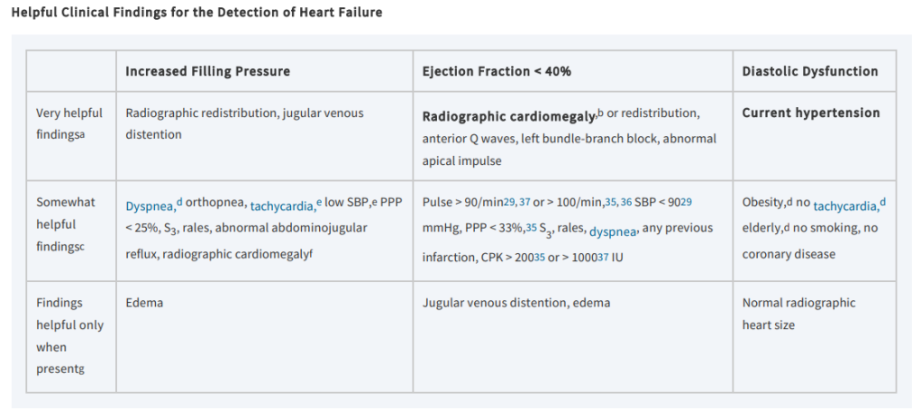
“Very helpful findings [for detection of heart failure] are radiographic redistribution and jugular venous distention. These findings, when used alone, only help when they are abnormal and so can confirm the presence of increased filling pressure in patients with known severe systolic dysfunction. Among patients referred for consideration of cardiac transplant with a high (73%) prevalence of increased filling pressure, radiographic redistribution indicates an 80% to 90% probability and jugular venous distention, an85% to 100% probability of increased filling pressure. The absence of either finding cannot rule out increased filling pressure. In patients with lesser probabilities of increased filling pressure, such as those without known severe systolic dysfunction, isolated findings may not be useful. Somewhat helpful findings include dyspnea and abnormal vital signs. Radiographic cardiomegaly is somewhat helpful but loses its specificity after the initial detection of increased filling pressure because it can be a permanent finding and not fluctuate with changes in filling pressure. Dependent edema is helpful only when present. Edema is highly specific for increased filling pressure, although it has poor sensitivity.” (Badgett)

(Badgett)

“Patients with heart failure can have decreased exercise tolerance with dyspnea, fatigue, generalized weakness, and fluid retention, with peripheral or abdominal swelling and possibly orthopnea. Patient history and physical examination are useful to evaluate for alternative or reversible causes. Nearly all patients with heart failure have dyspnea on exertion. However, heart failure accounts for only 30 percent of the causes of dyspnea in the primary care setting. The absence of dyspnea on exertion only slightly decreases the probability of systolic heart failure, and the presence of orthopnea or paroxysmal nocturnal dyspnea has a small effect in increasing the probability of heart failure (positive likelihood ratio [LR+] = 2.2 and 2.6).” (King)

Continue reading

EUH Morning Report: What are the sensitivity and specificity of the Kernig and Brudzinski sings for diagnosing adult meningitis? Part 1

The Bottom Line: “Since the overall accuracy of JAH and other clinical signs [of meningitis] is in poor to fair range, they do not have good performance alone in detection of meningitis. It seems that physicians should not solely rely on a single test or sign, and they should consider a collection of clinical signs and symptoms and history to have a better judgment. A lot of retrospective and prospective studies have shown that diagnosis of meningitis should be made according to the results of physical examination, history and CSF analysis.” (Ala)

In their 2018 prospective study, Ala et al. found that “that diagnostic value of JAH in diagnosis of meningitis in emergency department is higher than other clinical signs such as Kernig, Brudzinski and neck stiffness but the accuracy of all the mentioned signs is in poor to fair range (AUC 60 to 80). JAH had the highest sensitivity (84.4%) and Kernig and Brudzinski had the highest specificity (89.3% and 90.6%, respectively) among the evaluated signs.” (Ala)

(Ala)

This is congruent with the finds of prospective studies by Nakao et al. (2014) and Mofidi et al. (2017), that the Kernig and Brudzinski signs have low sensitivity but high specificity. And that along with Jolt Accentuation, the absence of the clinical signs of meningitis cannot rule out the diagnosis of meningitis.

“Although poorly sensitive, the physical examination findings of Kernig sign, Brudzinski sign, nuchal rigidity, vomiting, and rash were all relatively specific for both pleocytosis and moderate pleocytosis; however, because sensitivity was low for all of these features, LRs suggest that these findings are ultimately unhelpful. For the finding of moderate pleocytosis, however, high specificity resulted in potentially useful, although not diagnostic, LRs for a positive finding of jolt accentuation, Kernig sign, and Brudzinski sign.” (Nakao)

However, the “combination of Jolt accentuation, Kernig’s sign, and Brudzinski’s sign has 100% specificity and PPV and a very high LR+, so we can suppose a high possibility of meningitis for a patient with headache and fever and a positive result of all of 3 tests.” (Mofidi)

(Mofidi)

Ala, Alireza et al. “Accuracy of Neck stiffness, Kernig, Brudzinski, and Jolt Accentuation of Headache Signs in Early Detection of Meningitis.” Emergency (Tehran, Iran) vol. 6,1 (2018): e8. Free Full Text.

Nakao, Jolene H et al. “Jolt accentuation of headache and other clinical signs: poor predictors of meningitis in adults.” The American journal of emergency medicine vol. 32,1 (2014): 24-8. Full Text at Emory.

Mofidi, Mani et al. “Jolt accentuation and its value as a sign in diagnosis of meningitis in patients with fever and headache.” Turkish journal of emergency medicine vol. 17,1 29-31. 24 Nov. 2016. Free Full Text.

EUH Morning Report: What are the most useful clinical exam findings when diagnosing ascites?

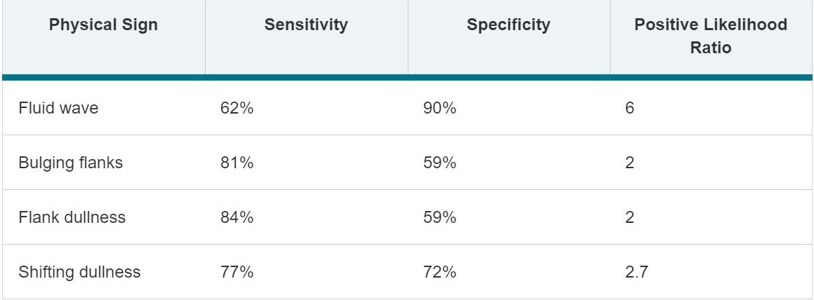
The Bottom Line (from The Rational Clinical Examination: Evidence-Based Clinical Diagnosis)

  1. The most useful findings for ruling out ascites are no history of ankle swelling or increased abdominal girth and the inability to demonstrate bulging flanks, flank dullness, or shifting dullness.
  2. The most powerful findings for making the diagnosis of ascites are a positive fluid wave result, shifting dullness, or peripheral edema.
  3. The puddle sign is difficult to perform, uncomfortable for patients, and not sensitive to small amounts of ascites. It should not be performed.
ascites

Continue reading

NEJM Videos in Clinical Medicine: Examination of the Neck Veins

Assavapokee T, Thadanipon K. Examination of the Neck Veins. N Engl J Med. 2020 Dec 10;383(24):e132. Full-text for Emory users.

Emory users, view video here.

“Examination of the neck veins can be easily performed in less than a minute and poses little risk to the patient. Direct examination of the neck veins, estimation of venous pressure, and performance of the abdominojugular reflux test are essential for evaluating right atrial pressure and estimating intravascular volume in patients with dyspnea, edema, or hypovolemia.”

EUH Morning Report: How do you define lymphadenopathy in relation to the lymph node region?

The  Bottom Line: Lymphadenopathy is defined as lymph nodes of abnormal size, generally > 1 cm, although definition of normal size range varies by lymph node regions and age of patient; for example:

  • jugulodigastric lymph nodes (often the largest of cervical lymph nodes) > 1.5 cm are considered abnormal
  • epitrochlear lymph nodes > 5 mm are considered abnormal
  • any palpable supraclavicular, popliteal, or iliac lymph nodes are considered abnormal
  • abdominal lymph nodes vary from 6-10 mm; retrocrural lymph nodes > 6 mm, retroperitoneal lymph nodes > 10 mm, and pelvic lymph nodes > 8-10 mm are considered abnormal
  • inguinal lymph nodes > 1.5 cm in diameter are considered abnormal

Continue reading

The Rational Clinical Examination: Heart Failure

Table 16-2. Helpful Clinical Findings for the Detection of Heart Failure.

Very helpful findingsa:

  • Increased Filling Pressure (IFP): Radiographic redistribution, jugular venous distention  Current hypertension
  • Ejection Fraction <40% (EF): Radiographic cardiomegalyb, or redistribution, anterior Q waves, left bundle-branch block, abnormal apical impulse
  • Diastolic Dysfunction (DD): Current hypertension

Somewhat helpful findingsc:

  • IFP: Dyspnea,d orthopnea, tachycardia,e low SBP,e PPP < 25%, S3, rales, abnormal abdominojugular reflux, radiographic cardiomegalyf
  • EF <40%: Pulse > 90/min or > 100/min, SBP < 9029 mmHg, PPP < 33%, S3, rales, dyspnea, any previous infarction, CPK > 20035 or > 100037 IU
  • DD: Obesity,d no tachycardia,d elderly,d no smoking, no coronary disease

Continue reading

EUH Morning Report: How do you perform a HINTS exam?

The Bottom Line:

  • The HINTS plus exam is a series of four bedside tests: head impulse testnystagmus, and test-of-skew, plus a bedside test for new hearing loss.
  • used to distinguish patients with continuous vertigo (lasting hours to days) and nystagmus who have a peripheral disorder (most likely vestibular neuritis) from those who have a central disorder (most likely cerebellar stroke).
  • do not perform HINTS test in patients with other syndromes besides acute vestibular syndrome, as may erroneously imply stroke in patients with benign positional vertigo (BPPV)

Continue reading

Krakow Conference: What are teaching points for JVP measurement and how to identify pulse abnormalities?

INSPECTION OF PRECORDIUM AND NECK (PG. 79)

  1. One must always inspect the chest wall for pulsations at the apex or in the precordium; this is best performed in left lateral decubital position.
  2. One must always inspect the right neck for presence and potential elevation of neck veins; best performed in supine position.
  3. Neck vein assessment provides information that further defines and delineates a problem; in and of itself it will not lead to a diagnosis.
  4. An elevated jugular venous pressure (JVP) is consistent with right ventricular dysfunction.
  5. The a wave corresponds to the atrial contraction and an S4. Atrial fibrillation has no specific a waves.
  6. The c wave is the most difficult to see.
  7. The v wave is an excellent wave to assess the tricuspid valve.
  8. Kussmaul’s sign is not specific to constrictive pericarditis. It is caused by right ventricular failure and infarction.
  9. Arm forward flexion and hepatojugular reflux are both complementary tests to the standard JVP .
  10. Central venous pressure (CVP) = JVP + 5. This is a more fastidious measure of neck vein height that effectively means the same as JVP.

From Cardiovascular Examination – Practice and Teaching in the Atlas of Adult Physical Diagnosis, edited by Dale Berg, and Katherine Worzala, Wolters Kluwer Health, 2005.


TYPES OF PULSE ABNORMALITIES: 

Pulse patterns

FIG. 16.9. Pulse abnormalities.

From Blood Vessels in the Seidel’s Guide to Physical Examination, edited by Jane W. Ball, Elsevier, 2019.